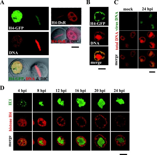
FIG. 1.
Chromatin marginalization is coupled with VS expansion. (A) BmN cells were transfected with plasmids expressing H4-GFP and H4-DsR, and the cells during interphase and mitotic phase were analyzed by confocal microscopy. H4-GFP-expressing cells were fixed at 24 h posttransfection and stained with a DNA-specific dye (PI) before the microscopic analysis. (B) Following transfection with a plasmid expressing H4-GFP, BmN cells were infected with a wild-type virus, fixed at 24 hpi, stained with a DNA-specific dye (PI), and analyzed by confocal microscopy. (C) BmN cells were mock infected or infected with a wild-type virus and fixed at 24 hpi. The fixed cells were hybridized with biotinylated virus DNA, stained with fluorescein-conjugated avidin and a DNA-specific dye (PI), and analyzed by confocal microscopy. (D) Following transfection with a plasmid expressing H4-DsR, BmN cells were infected with a recombinant virus expressing IE1-GFP and analyzed by confocal microscopy at the indicated time points. Bars, 10 μm.

