FIG. 3.
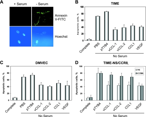
Viral chemokine-mediated protection of endothelial cells from starvation-induced apoptosis. (A) Examples of Annexin V-FITC staining to detect apoptosis in TIME cells deprived of serum for 48 h. (B) Quantified data from an experiment in which serum-starved cultures were supplemented with recombinant vCCL-1, vCCL-2, CCL1, or VEGF, or to which PBS or “pTYB4-extract” (negative controls) was added. (C) An analogous experiment was carried out in primary endothelial cells, DMVEC. (D) Similar analysis of TIME-CCR8i and TIME-NS (control) cells demonstrated the dependence on CCR8 for the protective effects of vCCL-1, vCCL-2, and CCL1, but not of non-CCR8 ligand VEGF. (Maximally effective chemokine and VEGF concentrations of 250 ng/ml and 10 ng/ml, respectively, were used for experiments B to D.)

