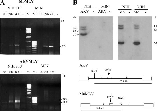
FIG. 3.
Restriction of AKV MLV in M. minutoides cells occurs before reverse transcription. (A) Hirt DNA extracts prepared at several time points after virus infection were used to detect 2-LTR viral DNA circles in infected NIH 3T3 and M. minutoides (MIN) cells. PCR identified a doublet in the NIH 3T3 cells that sequencing confirmed to represent LTRs with one or two enhancer copies. Marker lanes are labeled M. Dashes represent uninfected cells. (B) Southern blot analysis of SacII-cut Hirt DNA extracted 3 days after virus infection using a pol-specific hybridization probe. NIH 3T3 and MIN cells were infected with AKV MLV or MoMLV. Dashes represent uninfected cells. Diagrams at the bottom show the positions of the SacII sites and the hybridization probe in the two virus genomes.

