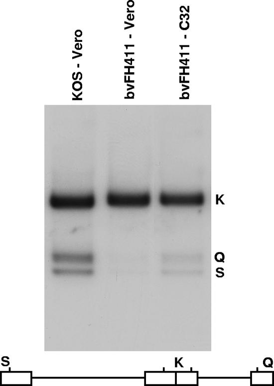
FIG. 4.
Processing of viral DNA. Vero or C32 cells were infected with the indicated virus at an MOI of 5 PFU per cell. Total infected-cell DNA was isolated at 16 h postinfection, digested with BamHI, and subjected to Southern blot analysis using the BamHI K joint fragment (32P labeled) as a probe. Scanned images of the autoradiograph obtained from the Southern blots are shown. The locations of the BamHI K joint fragment and the two end fragments, BamHI-Q and -S, in the HSV-1 genome are shown at the bottom.

