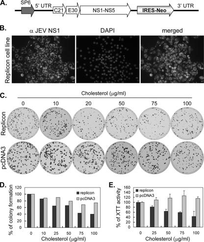
FIG. 9.
JEV replicon cell growth is slightly reduced by cholesterol treatment. (A) Schematic representation of the JEV replicon construct. SP6, SP6 promoter. “5′ UTR” and “3′ UTR” represent the 5′ and 3′ UTRs, respectively. C21 corresponds to the first 21 amino acids of JEV core protein, and E30 corresponds to the last 30 amino acids of JEV E protein. NS1-NS5 corresponds to the sequence coding for the JEV nonstructural proteins. IRES-Neo represents a sequence of an IRES of encephalomyocarditis virus followed by a neomycin resistance gene (Neo). (B) Establishment of a JEV replicon cell line. BHK-21 cells were transfected with in vitro-transcribed replicon RNA and selected with G418. The G418-resistant cells were stained with anti-JEV NS1 plus fluorescein isothiocyanate-conjugated secondary antibody and DAPI. (C) Colony formation of JEV replicon cells in the presence of cholesterol. The replicon cells and the pcDNA3-transfected BHK-21 cells were cultured with a G418-containing agarose overlay plus different concentrations of cholesterol as listed at the top of the panel. Colony formation was assessed by crystal violet staining after 1 week of incubation. (D) Replicon cell colony formation was slightly reduced by cholesterol treatment. The crystal violet-stained cells were lysed, and the level of optical density absorbance at an optical density of 570 nm was determined. The data are derived from the average results obtained with four plates for each experimental group. (E) JEV replicon cell growth was reduced by cholesterol treatment. The indicated cells were cultured with G418 plus cholesterol at different concentrations for 24 h before the cell growth was monitored by XTT assays. The averages and standard deviations of the results obtained with triplicate samples are shown.

