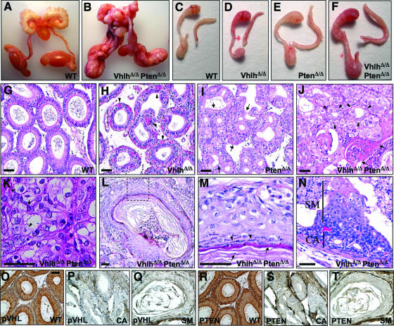
FIG. 3.
Phenotypic analysis of Vhlh and Pten deletion in the male genital tract. (A and B) Genital tracts of 14-week-old male wild-type (A) and VhlhΔ/Δ PtenΔ/Δ (B) mice. (C to F) Epididymides and vasa deferentia of 8-week-old wild-type (C), VhlhΔ/Δ (D), PtenΔ/Δ (E), and VhlhΔ/Δ PtenΔ/Δ (F) mice. (G to J) Hematoxylin- and eosin-stained sections of epididymides of 8-week-old wild-type (G), VhlhΔ/Δ (H), PtenΔ/Δ (I), and VhlhΔ/Δ PtenΔ/Δ (J) mice. Arrowheads in panel H point to blood vessels, regions between arrows in panel I highlight regions of hyperplasia, arrowheads in panel J point to adenoid structures in a region of cystadenoma, and arrows in panel J point to a region of squamous differentiation. (K) Region of cystadenoma in the epididymis of a 14-week-old VhlhΔ/Δ PtenΔ/Δ mouse with examples of clear cells shown by arrowheads. (L) Epididymal tubule showing squamous metaplasia in a 14-week-old VhlhΔ/Δ PtenΔ/Δ mouse. (M) Zoom of the dotted boxed region in panel L. (N) Early lesion in an epididymal tubule of an 8-week-old VhlhΔ/Δ PtenΔ/Δ mouse. A region that appears to be a precursor of cystadenoma is labeled CA, and a region of cells undergoing squamous metaplasia is labeled SM. (O to Q) Immunohistochemical staining using an antibody against pVHL in epididymal tubules from a wild-type (O) mouse or from regions of cystadenoma (P) and squamous metaplasia (Q) from a VhlhΔ/Δ PtenΔ/Δ mouse. (R to T) Immunohistochemical staining using an antibody against Pten in epididymal tubules from a wild-type (R) mouse or from regions of cystadenoma (S) and squamous metaplasia (T) from a VhlhΔ/Δ PtenΔ/Δ mouse. Panels O to T are at the same magnification. Bars, 50 μm.

