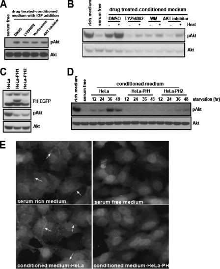
FIG. 2.
The PI3K-Akt signaling pathway mediates the secretion of Akt-activating activity. (A) HeLa cells were cultured in serum-free medium for 24 h in the presence of dimethyl sulfoxide (DMSO), LY294002 (5 μM), wortmannin (WM, 0.1 μM), or Akt inhibitor (3.6 μM). The CM was filtered through a Centriprep-10K column to remove the residual chemicals (see Materials and Methods). The filtered CM was mixed with IGF-1 (1 ng/ml) and tested for the removal of residual drug. (B) Each CM sample from the experiment described in the legend to panel A was untreated or heat treated, and its ability to activate Akt was assayed in HeLa cells. (C) The whole-cell lysates of HeLa cells or HeLa-PH cells grown in serum-rich medium were analyzed for phospho-Akt level. (D) The serum-free CM from HeLa, HeLa-PH1, or HeLa-PH2 cells was collected at 12-h intervals and tested for its ability to activate Akt. (E) The CM from HeLa or HeLa-PH cells was added to HeLa-PH cells that had been serum starved for 12 h. After the HeLa-PH cells were incubated for 30 min, they were fixed in 3% paraformaldehyde, and the membrane translocation of PH-EGFP was examined using fluorescence microscopy (20× objective). Arrows indicate the membrane translocation of PH-EGFP.

