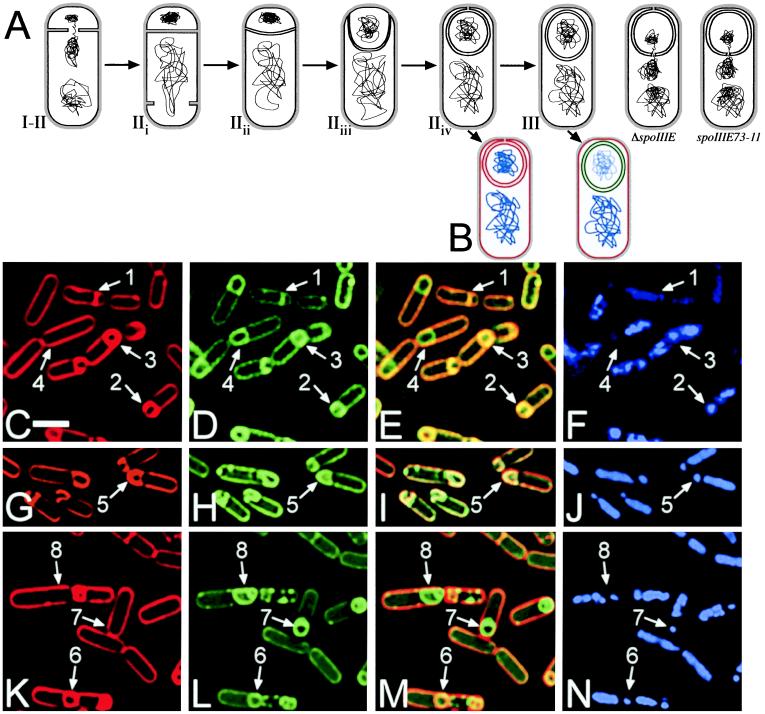
Figure 1.
(A) Stages of engulfment, showing completion of chromosome translocation and polar septation (I–IIi), early engulfment (IIii), membrane movement across the cell pole (IIiii), and a stage of engulfment newly defined here, mother cell membrane fusion (IIiv). The two cells at the right illustrate the proposed defects in ΔspoIIIE and spoIIIE73-11. (B) Schematic of the membrane fusion assay. Red lines represent membranes stained with FM 4-64 and MTG, green lines indicate membranes stained with MTG only, and blue lines show DAPI staining of chromosomes. (C–N) Sporangia of PY79 (C–F), KP141 (G–J), and KP541 (K–N) stained with FM 4-64 (C, G, and K), MTG (D, H, and L), and DAPI (F, J, and N) 3 hr after the initiation of sporulation (t3); E, I, and M are an overlay of FM 4-64 and MTG staining. Arrows 1 and 2 show cells at stage IIi and IIii–iii respectively. Arrows 3, 5, and 6 point to cells at stage IIiv which have completed membrane migration but have not yet completed membrane fusion as shown by the FM 4-64 (C, G, and K) and MTG (D, H, and L) staining. Finally, arrows 4, 7, and 8 indicate sporangia that have completed membrane movement and fusion; only the mother cell membranes stain with FM 4-64 (C and K), but MTG stains both the forespore and mother cell membranes (D and L). The reduced forespore chromosome content of spoIIIE mutants can be seen in N and J (arrows 5–8). F shows that after the completion of membrane fusion the forespore chromosome stains less intensely with DAPI in wild-type sporangia (compare arrows 3 and 4). (White bar in C is 2 μm.)

