Figure 4.
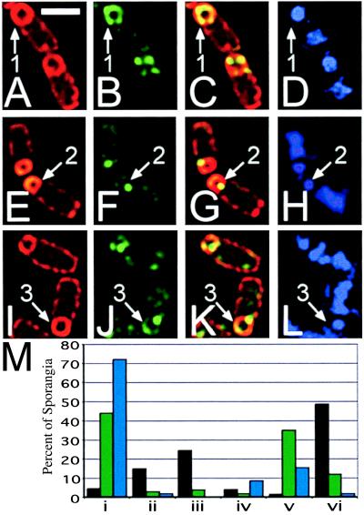
Cells of KP542 (wild type; A–D), KP538 (spoIIIE36; E–H), and KP537 (spoIIIE73-11; I–L) immunostained with anti-GFP antibodies (B, C, F, G, J, and K, green) FM 4-64 (A, B, E, F, I, and J, red) and DAPI (D, H, and L) at t3. The predominant staining pattern for each strain is shown; fields are shown in supplemental Fig. 7 at www.pnas.org. Arrow 1 shows wild-type SpoIIIE-GFP completely surrounding the forespore of an engulfed sporangium. Arrow 2 shows the predominant septal immunostaining pattern of SpoIIIE36-GFP. Arrow 3 indicates the localization of SpoIIIE73-11-GFP to a septal focus and in a crescent at the forespore pole. (White bar in A is 2 μm.) (M) Scoring of SpoIIIE-GFP localization in fully engulfed sporangia, SpoIIIE-GFP (black bars), SpoIIIE73-11-GFP (green bars), SpoIIIE36-GFP (blue bars). i, sporangia with a single focus of fluorescence in the middle of the polar septum; ii, sporangia with a single focus of fluorescence on the side of the forespore; iii, sporangia with a single focus of fluorescence at the cell pole; iv, sporangia with two foci of fluorescence, one at the polar septum and one on the side of the forespore; v, sporangia with two foci of fluorescence, one at the septum and one at the cell pole; vi, sporangia with delocalized fluorescence or with more than two foci. At least 100 fully engulfed sporangia were scored for each strain; values represent percentages of scored sporangia. More detailed scoring is shown in supplemental Fig. 7 at www.pnas.org.

