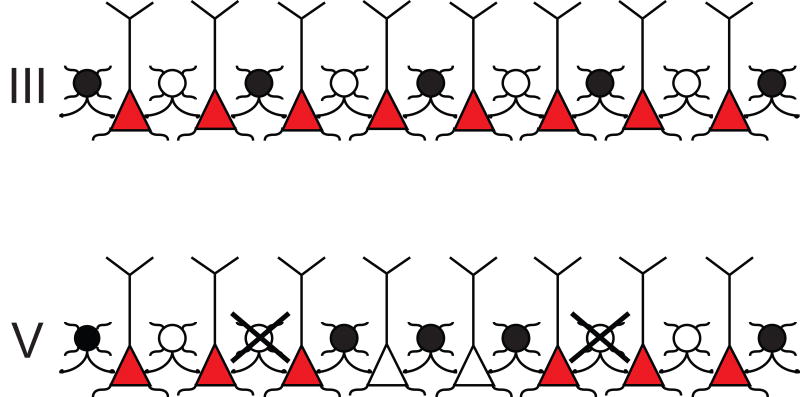
Figure 4.
A schematic diagram that summarizes the potential lamina-specific focusing of pyramidal cells by PV+/GABA inhibition in the PrL cortex after the repeated AMPH paradigm. Filled circles (•) indicate PV+ interneurons that contain Fos, whereas open circles (○) represent interneurons that show no Fos activation. Interneurons that have lost their PV immunoreactivity are illustrated as crossed (X) open circles. Red and empty triangles represent pyramidal neurons. It is well known that the PV+ interneurons provide a direct inhibition of pyramidal cells by synapsing onto their cell bodies or proximal axonal shafts in the same cortical layer (Hendry et al., 1983; DeFelipe et al., 1989; Kawaguchi and Kubota, 1993). Following repeated administration of AMPH, the density of co-labeled interneurons is unchanged in layers III and V. However, the proportion of activated PV+ neurons is increased in layer V due to the loss of PV-immunoreactivity. These data support a model in which the pattern of PV+ activation will presumably synchronize outflow from this layer, by silencing some pyramidal cells (empty triangles) and leaving others activated (red triangles).

