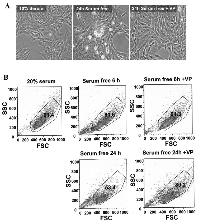
Fig. 1.
Serum starvation induced cell death in H32 hypothalamic cells. (A) Light microscopy images of H32 hypothalamic cells following incubation with 10% serum, or serum starvation for 24h in the presence or in the absence of 10nM VP. (B) Flow cytometry analysis of the cell size (Forward Scatter [FSC]) and granularity (Side Scatter [SSC]) of H32 hypothalamic cells following incubation for 6 or 24 h with 20% serum, serum-free medium or serum-free medium plus 10nM VP. The gated population represents the percentage of living cells. Each plot is representative of three experiments with similar results.

