Fig. 6.
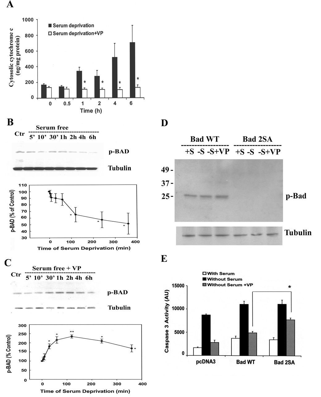
VP induces the phosphorylation of pro-apoptotic protein, BAD, and prevents the release of cytochrome c form mitochondria. (A) The time course of cytosolic cytochrome c in the presence and the absence of 10nM VP. Cytochrome c levels were measured using an ELISA Kit as described in Methods. Bars represent the mean ± S.E.M of three experiments conducted in duplicate. * p< 0.05 and ** p< 0.01, significantly different form the corresponding serum deprivation group. Time course of BAD phosphorylation in the absence (B) or in the presence of 10 nM VP (C). Phosphorylation of BAD (p-Bad Ser112) was examined by Western blotting. Data are expressed as the mean ± S.E.M of three experiments. * p< 0.05, significantly different form the control group. PCDNA3 or Bad wild type (BAD WT) or Bad dominant negative mutant (Bad 2SA) was transfected into H32 cells. 24h after transfection, cells were incubated in 20% serum(+S) or serum-free medium(−S), or serum-free medium with 10nM VP(−S+VP). (D) Phosphorylation of BAD (Ser112) was examined by Western blotting, and (E) caspase-3 activity was determined as described in “Materials and Methods”. The bars represent the mean ± S.E.M of three experiments conducted in duplicate. * p< 0.05, compared to Bad WT Without Serum+VP group. .

