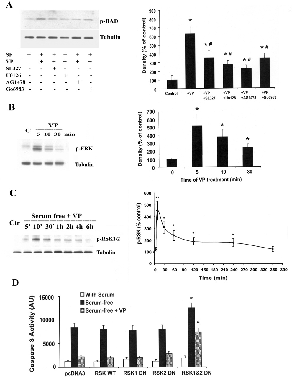
Fig. 7.
Upstream signaling pathways involved in VP-induced BAD phosphorylation. (A) Effect of the ERK-MAP kinase inhibitors, SL327 (1µM) and UO126 (1µM), the EGF receptor inhibitor, AG1478 (100 µM) and the PKC inhibitor Go6983 (1µM) on VP-induced BAD phosphorylation. H32 cell incubated in serum-free medium for 2 h (control) were pre-incubated with the inhibitors for 30 min before addition of 10nM VP. Immunoblot analysis of phosphorylation of BAD (Ser112) was conducted as described in Method. For measurement of the effects of VP on ERK phosphorylation (B) and RSK phosphorylation (C), H32 hypothalamic cells were incubated in serum-free medium (SF) for 2h (control), before addition of 10 M VP. Cell extracts for Western blotting analysis of p-ERK and p-RSK were prepared at the time points indicated in the graphs. Data represent the mean and SEM of the results obtained in three experiments. * p< 0.05 and p<0.01, compared to serum-free (control) group, # p< 0.05 compared to serum-free + VP group. (D) PCDNA3 or RSK wild type (RSK WT) or RSK dominant negative mutants (RSK 1, RSK2 or RSK1&2) were transfected into H32 cells. 24h after treansfection, cells were incubated in 20% serum (With Serum), or serum-free medium (Serum-free), or serum-free medium with 10nM VP (Serum-free+VP). Caspase-3 activity was determined. The bars represent the mean ± S.E.M of three experiments conducted in duplicate. * p< 0.05, compared to RSK WT serum-free group, # p< 0.05 compared to RSK WT serum-free + VP group‥

