Figure 1.
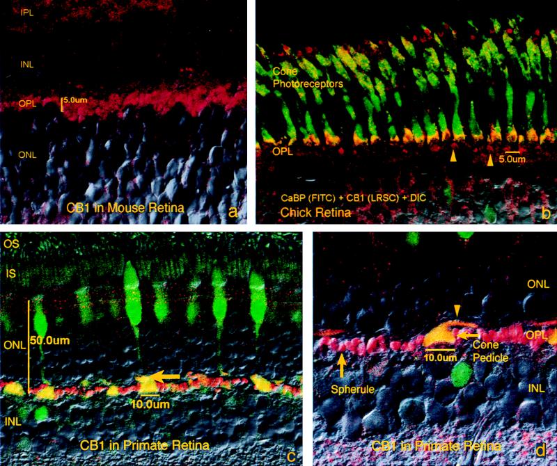
CB1 is present in cone pedicles and rod spherules. Combined fluorescence and Nomarski micrograph. Photoreceptor inner segments (IS), outer nuclear layer (ONL), OPL, INL, IPL, and GCL. (a) CB1 labeling is found in the OPL of mouse retina in what probably correspond to spherules in the rod-dominated retina of the mouse. (b) Image of calbindin double-labeled with CB1 in chick retina. Cone photoreceptors are labeled in green (calbindin, CaBP). CB1 labeling is shown in red. Yellow double-labeling occurs within the pedicles of cone photoreceptors. Arrowheads indicate pedicle labeling in layers two and three of the chick OPL. (c) Labeling of the primate OPL shows very distinctly labeled pedicles (yellow, arrow) and spherules. (d) Higher magnification view of monkey OPL shows a calbindin/CB1 double-labeled cone pedicle as well as CB1 labeled rod spherules. Pedicle stalk is indicated by arrowhead. Magnifications: (a and b) ×900; (c) ×600; (d) ×950.

