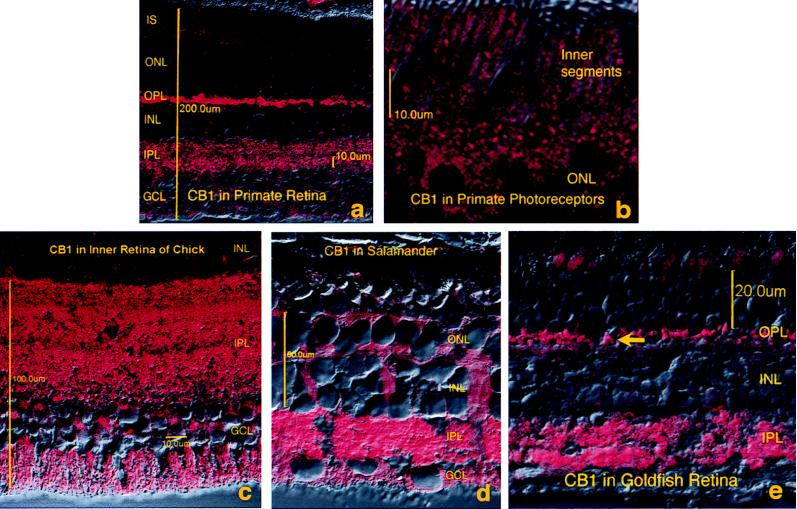
Figure 3.
CB1 labeling is found in a similar pattern in a wide range of vertebrate retinas. Combined fluorescence and Nomarski micrographs of transverse cross-sections of the retina. Photoreceptor inner segments (IS), outer nuclear layer (ONL), OPL, INL, IPL, GCL. (a) Overview of monkey retina, with pronounced labeling in OPL and IPL. (b) Higher magnification view of the photoreceptor layer of the primate retina, with CB1 labeling in the inner segments. (c) IPL of chick, showing robust labeling with some stratification. Somatic labeling in some ganglion and amacrine cells is also visible, as is labeling in ganglion cell axons. (d) Overview of retina of the tiger salamander. Labeling is present in OPL, INL, IPL, and GCL as well as ganglion cell axons. (e) Overall view of goldfish retina showing all layers. Labeling corresponds to rod spherules and cone pedicles (arrow) in the OPL. Labeling in the IPL is patchy because of tissue folding. Labeling in the photoreceptors is caused by autofluorescence. Magnifications: (a) ×230; (b) ×1,100; (c) ×450; (d) ×410; (e) ×625.

