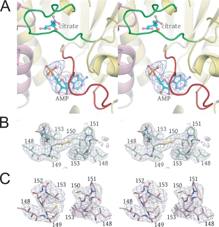
FIGURE 5.
Difference and annealed omit electron density superimposed on key regions of IDH. A, the AMP-binding site is revealed by Fo - Fc electron density contoured at 3σ after rigid body refinement of the citrate-bound IDH structure against the 4.3 Å citrate + AMP diffraction data. The orientation is approximately the same as that shown in Fig. 4, A and B. B, annealed omit map contoured at 3σ calculated for the unliganded IDH structure in which IDH2 residues 148-153 were removed from the phase calculations. The electron density reveals an oxidized disulfide bond between symmetry-related IDH2 Cys-150 residues. C, annealed omit map contoured at 4σ calculated for the citrate-bound IDH structure in which IDH2 residues 148-153 were removed from the phase calculations. The electron density reveals a reduced disulfide bond between symmetry-related IDH2 Cys-150 residues (see “Results”).

