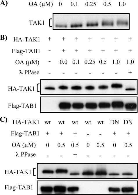
FIGURE 1.
Inhibition of Ser/Thr protein phosphatases by OA induces TAK1 phosphorylation. A, OA treatment increases mobility shift in endogenous TAK1. MMC grown to subconfluence were treated with increasing concentrations of OA for 6 h as indicated. Cell lysates were subjected to Western blot analysis with anti-TAK1 antibody. B, OA treatment enhances TAK1 phosphorylation induced by coexpression of TAK1 and TAB1. MMC transfected with expression vector encoding HA-TAK1 alone or together with FLAG-TAB1 were treated with increasing concentrations of OA for 6 h, as indicated. Expression of HA-TAK1 and FLAG-TAB1 was determined by Western blot analysis with anti-HA and anti-FLAG antibodies, respectively. Four hundred units of λ protein phosphatase (λ PPase) were added to cell lysates and incubated for 30 min at 30 °C to dephosphorylate TAK1. C, TAK1 kinase activity is required for its phosphorylation. Wild-type HA-TAK1 (wt) or kinase-deficient mutant of HA-TAK1 (DN) was coexpressed with FLAG-TAB1 in MMC, as indicated, and treated with 0.5 μm OA for 6 h. Western blot analysis and treatment with λ PPase were performed as described in B.

