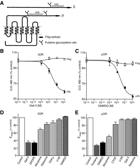
FIGURE 1.
Agonist-induced conformational changes in the N terminus of μOR and δOR. A, schematic representation of μOR and δOR. The FLAG epitope (black ellipse), putative N-linked glycosylation sites (branches), and amino acid sequences used for the generation of antigenic peptides (black lines) are indicated. B and C, CHO cells stably expressing δOR (C) or μOR (B) were treated with indicated doses of deltorphin II or DAMGO in isotonic HEPES buffer, pH 7.4 and probed with 5A2, 4D6, and FLAG antibodies by ELISA as described under “Experimental Procedures.” Absorbance obtained with cells not treated with agonist (1.27 ± 0.04 for 5A2, 1.44 ± 0.02 for 4D6, 1.03 ± 0.01 for FLAG in CHOμOR and 1.15 ± 0.07 for FLAG in CHOδOR) following subtraction of signal with only secondary antibody were taken as 100%. Results are mean ± S.E. of three experiments in triplicate. D and E, CHO cells stably expressing δOR (D) or μOR (E) were treated without or with 1 μm different ligands in 50 mm Tris-Cl buffer, pH 7.5 and probed with μOR (4D6) and δOR (5A2) mAbs by ELISA as described under “Experimental Procedures.” Data with vehicle-treated cells were taken as 100%. Results are mean ± S.E. of three experiments in triplicate. **, p < 0.01 Dunnett's test.

