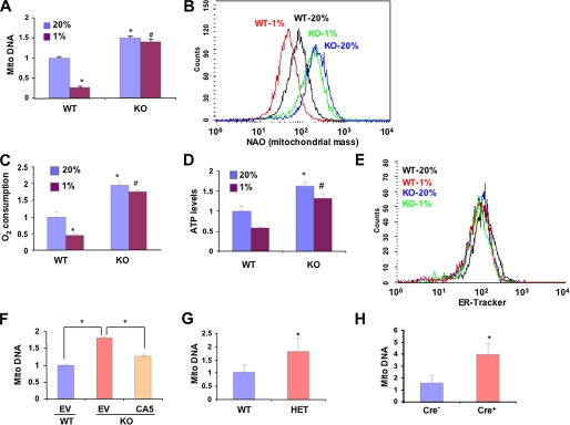
FIGURE 1.
Regulation of mitochondrial mass and respiration by HIF-1 ex vivo and in vivo. A, the ratio of mitochondrial:nuclear DNA was determined by quantitative real-time PCR in wild type (WT) and Hif1a-/- (KO) MEFs exposed to 20 or 1% O2 for 48 h and normalized to the results obtained for WT cells at 20% O2. Mean values are shown (±S.E.). *, p < 0.05 by Student's t test compared with WT MEFs at 20% O2;#, p < 0.05 compared with WT MEFs at 1% O2. B, WT and KO MEFs were exposed to 20 or 1% O2 for 48 h. Equal numbers of cells were stained with nonyl acridine orange (NAO) and analyzed by flow cytometry to measure mitochondrial mass. C and D, O2 consumption (C) and ATP levels (D) were measured in WT and KO MEFs exposed to 20 or 1% O2 for 48 h and normalized to the results obtained for WT MEFs at 20% O2. Mean values are shown (±S.E.). *, p < 0.05 by Student's t test compared with WT MEFs at 20% O2;#, p < 0.05 compared with WT MEFs at 1% O2. E, WT and KO MEFs were exposed to 20 or 1% O2 for 48 h. Equal numbers of cells were stained with ER-Tracker and analyzed by flow cytometry to measure endoplasmic reticulum mass. F, WT and KO MEFs were transduced with empty retroviral vector (EV) or vector encoding constitutively active HIF-1α (CA5). After 3 days the ratio of mitochondrial:nuclear DNA was determined. Mean values are shown (±S.E.). *, p < 0.05 for indicated comparison. G and H, DNA was isolated from lungs of WT and Hif1a+/- HIF-1α-HET littermate mice (G) or Arntflox/flox HIF-1β-conditional-knock-out mice that were either transgenic (Cre+) or non-transgenic (Cre-) for Tie2-Cre (H). The ratio of mitochondrial: nuclear DNA was determined by real-time PCR and normalized to the results obtained for WT (G) or Cre- (H) mice. *, mean (± S.E., n = 3) that is significantly different from WT or Cre-.

