FIGURE 9.
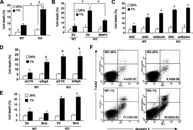
Protective effect of HIF-1/BNIP3/Beclin/Atg6-induced autophagy in hypoxic cells. A, B, C, D, and E, the indicated MEF subclones were cultured at 20 or 1% O2 for 48 h, and the number of dead cells as a percentage of total cell number was determined by trypan blue staining. Mean data (±S.E.) are shown. *, p < 0.05 by Student's t test compared with the control WT MEF subclone in the first column of each bar graph. #, p < 0.05 for indicated comparison (A and B) or compared with WT-SNC (C), siCTR (D), or WT-EV (E) at 1% O2. F, MEFs were cultured at 20 or 1% O2 for 48 h and then incubated with 7-AAD and phosphatidylethanolamine-labeled anti-annexin V antibody for flow cytometric analysis of apoptosis. The percentage (mean ± S.E.) of annexin+/7-AAD- cells are shown.

