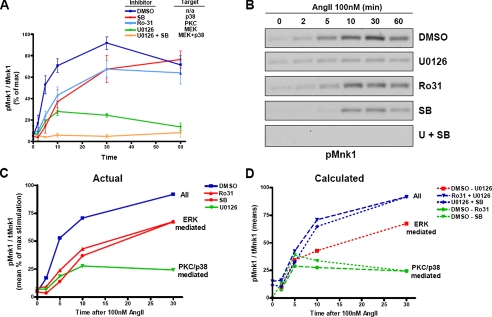
FIGURE 1.
Sensitivity of AngII-stimulated pMnk1 activation to kinase inhibitors. A, HEK-293 cells stably expressing HA-AT1AR and Mnk1 were serum-starved for 16 h, pretreated with vehicle Me2SO (DMSO) or the indicated kinase inhibitor for 30 min, and then stimulated with 100 nm AngII for the indicated times. Western blots for phospho-Mnk1 and tMnk1 were performed. The percentage of pMnk1/tMnk1 was calculated for each data point and plotted as a percentage of maximal activity (Me2SO-treated cells at 30 min) ± S.E. These data represent the average ± S.E. of seven independent experiments. B, representative pMnk1 Western blots. C, means of individual data points (as a percent of maximal stimulation) up to 30 min from experiments performed in A. D, calculations based on addition or subtraction of means from A as shown in the legend. Solid lines in C represent actual data; dashed lines in D represent mathematically calculated values.

