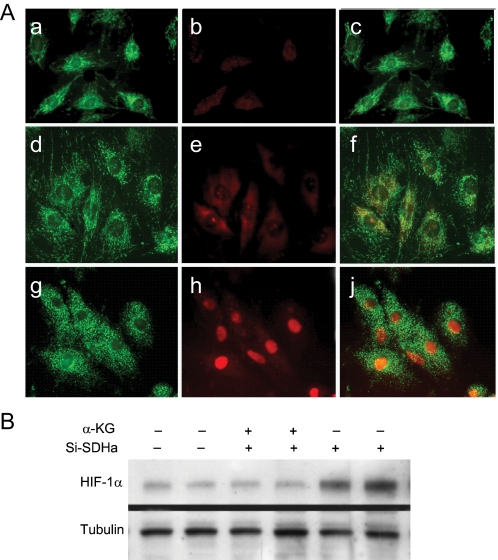
FIGURE 6.
Prevention of HIF-1α nuclear translocation by α-ketoglutarate in SDHa-silenced H9c2 cells. A, confocal images of control H9c2 cells stained with MitoTracker Green (panel a) and Cy3 immunostaining for HIF-1α (panel b, red) and overlay (panel c); or SDHa-silenced cells with α-KG and MitoTracker Green (panel d) and HIF-1α (panel e, red) and overlay (panel f); or SDHa-silencing without α-KG and MitoTracker Green (panel g) and HIF-1α (panel h, red) and overlay (panel j). B, Western blot of nuclear extracts (20 μg) using antibodies to HIF-1α and tubulin is shown.

