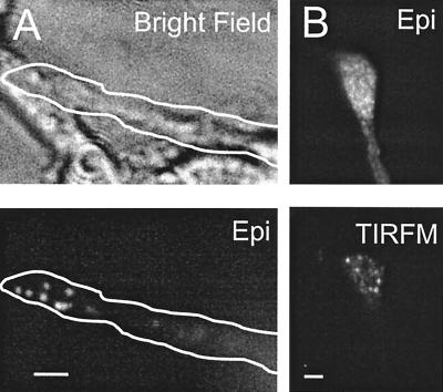
Figure 1.
Single GFP-labeled secretory vesicles in neuronal growth cones. (A) Cells were treated with 1 μM muristerone to activate the inducible expression construct before imaging. (Upper) Bright-field view of neurites illuminated with a Hoffman condenser. (Lower) Epifluorescence image shows that single secretory vesicles labeled with proANF-Emd are evident in one growth cone. Outline shows transfected neurite. (B Upper) Conventional epifluorescence image of a growth cone expressing the constitutive proANF-Emd construct. Note that it is difficult to resolve individual vesicles. (Lower) Single secretory vesicles are evident in the TIRFM image of the same growth cone. (Bar = 2 μm.)

