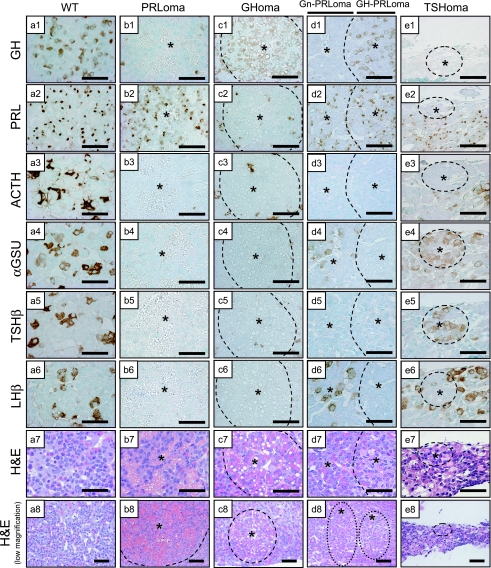
Fig. 2.
Immunocharacterization of pituitary hormones in Prop1 transgenic adenomas. Light microscopy of coronal sections of a wild-type mouse pituitary (WT; a) and Prop1 Tg mice including PRLoma (b), Prop1 Tg including GHoma (c), Gn-PRLoma/GH-PRLoma (d) and small TSHoma (e). Immunostaining of GH, PRL, ACTH, αGSU, TSHβ, LHβ and H&E stain. All sections were stained by methyl green nuclear stain. Asterisk: adenoma region. Bars=50 µm.

