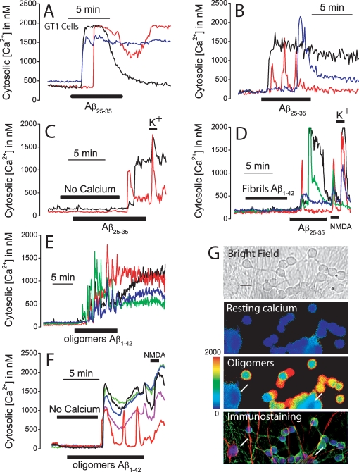
Figure 1. Aβ1–42 oligomers but not fibrils induce Ca2+ influx in neurons.
A–B. GT1 neural cells and cerebellar granule cells were loaded with fura2/AM and subjected to calcium imaging. Traces show the effects of Aβ25–35 (20 µM) on [Ca2+]cyt in three representative GT1 neural cells (A) and cerebellar granule cells (B). Data representative of 141–233 cells studied in 4 and 3 independent experiments, respectively. C. The increase in [Ca2+]cyt induced by Aβ25–35 (20 µM) in cerebellar granule cells is abolished in medium lacking extracellular Ca2+ (No calcium). Addition of normal medium containing 1 mM Ca2+ restored the response to Aβ25–35. High K+ medium (150 mM K+) induced a further increase in [Ca2+]cyt. Traces correspond to two individual cells representative of n = 98 cells, 2 experiments). D. Aβ1–42 fibrils (2 µM) induced little or no [Ca2+]cyt increase in cerebellar granule cells. The same cells responded Aβ25–35 (20 µM), N-methyl D-aspartate (100 µM NMDA) and high K+ medium (150 mM K+). (Traces correspond to four individual cells representative of n = 90 cells 3 experiments). E. Aβ1–42 oligomers (500 nM) induced a large and sustained increase in [Ca2+]cyt in cerebellar granules cells. Traces correspond to four individual cells representative of n = 404 cells 5 experiments). F. The effects of Aβ1–42 oligomers (500 nM) are inhibited in medium lacking extracellular Ca2+ (No calcium). Perfusion of medium containing Ca2+ restored the response to Aβ oligomers. Cells also responded to 100 µM NMDA (recordings correspond to 5 individual cells representative of n = 283 cells, 3 experiments). G. Cerebellar granule cells were subjected to calcium imaging and then identified by double immunocytochemistry. Pictures show a bright field image (scale bar represents 10 µm) and [Ca2+]cyt levels before (resting calcium) and after treatment with Aβ1–42 oligomers (oligomers) coded in pseudocolor (pseudocolor bar from 0 to 2000 nM shown at left) and immunostaining of the cells. Glial cells (arrows) are coded in red, neurons are coded in green and nuclei are coded in blue. Only neurons responded to Aβ oligomers with a rise in [Ca2+]cyt. Data representative of 249 cells, 3 experiments.

