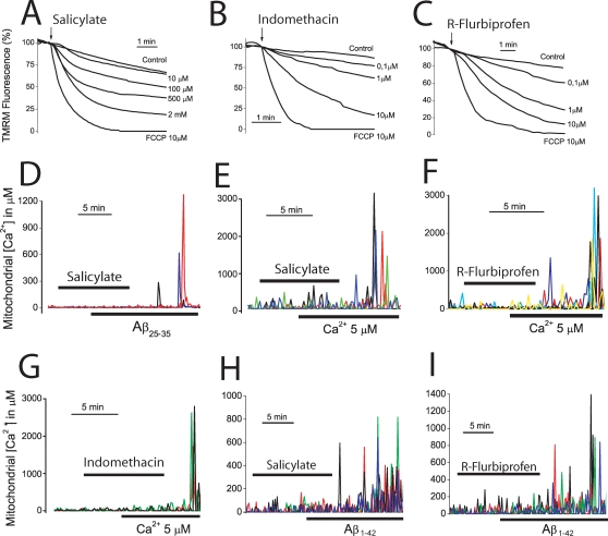
Figure 6. NSAIDs depolarize mitochondria and inhibit mitochondrial Ca2+ uptake.
A–C. Cerebellar granule cells were stained with 10 nM TMRE and subjected to fluorescence microscopy for monitoring changes in mitochondrial potential. Each trace correspond to averaged, normalized fluorescence values of 34–42 cells treated (arrow) with vehicle (control) or different concentrations of salicylate (A, 10 µM to 2 mM), indomethacin (B, 0.1 to 10 µM), R-flurbiprofen (C, 0.1 to 10 µM). FCCP 10 µM was also added to compare with conditions of collapse of the mitochondrial potential. Each series of recordings is representative of at least 3 experiments. D–F. Cerebellar granule cells were transfected with the mGA plasmid and subjected to bioluminescence imaging for monitoring [Ca2+]mit. Salicylate (D, 100 µM) inhibits the [Ca2+]mit induced by Aβ25–35 (20 µM, n = 43 cells, 3 experiments). In some experiments, cerebellar granule cells expressing mGA were permeabilized in intracellular medium containing 200 nM Ca2+ (see methods) and treated with salicylate 100 µM (E), R-flurbiprofen 1 µM (F) or indomethacin 1 µM (G) before being stimulated with the same intracellular medium containing 5 µM Ca2+ to stimulate mitochondrial Ca2+ uptake (Data from 188 cells studied in 6 independent experiments). H,I. Salicylate 100 µM and R-flurbiprofen 1 µM also inhibits the increase in [Ca2+]mit induced by Aβ1–42 oligomers (500 nM). Data from n = 35 and 30 cells respectively, studied in 3 independent experiments for each drug.

