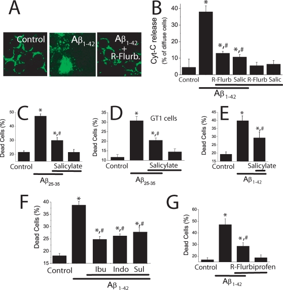
Figure 8. NSAIDs inhibit cytochrome c release and cell death induced by Aβ1–42 oligomers.
A,B. Cerebellar granule cells were treated with Aβ1–42 (500 nM) for 72 h with or without 1 µM R-flurbiprofen or 100 µM salicylate and fixed for analysis of cytochrome c location using confocal microscopy. A. Control cells showed a punctate distribution of cytochrome c. Scale bar represents 10 µm. Aβ1–42 oligomers-treated cells show a more diffuse pattern of cytochrome c whereas cells treated with Aβ1–42 oligomers plus R-flurbiprofen show a punctate pattern similar to control cells. B. Bars show the relative abundance (%) of cells showing diffuse immunostaining in control cells, cells treated with Aβ1–42 oligomers and cells treated with Aβ1–42 oligomers plus 1 µM R-flurbiprofen or 100 µM salicylate (B). *p<0 05 vs control; #p<0 05 vs Aβ; data from at least 3 independent experiments. Salicylate 100 µM inhibits cell death induced by Aβ25–35 (20 µM) in cerebellar granule cells (C) and GT1 cells (D). Salicylate 100 µM also inhibits cell death induced by Aβ1–42 oligomers (500 nM) in cerebellar granule cells (E). (*p<0 05 vs control; #p<0 05 vs Aβ. n = 3). F. Ibuprofen (Ibu), Indomethacin (Indo), and sulindac sulfide (Sul), all tested at 1 µM, inhibit cell death induced by Aβ1–42 oligomers (500 nM) in cerebellar granule cells. G. R-Flurbiprofen 1 µM also inhibits cell death induced by Aβ1–42 oligomers (500 nM). *p<0 05 vs. control; #p<0 05 vs Aβ. All data are representative of 3 experiments.

