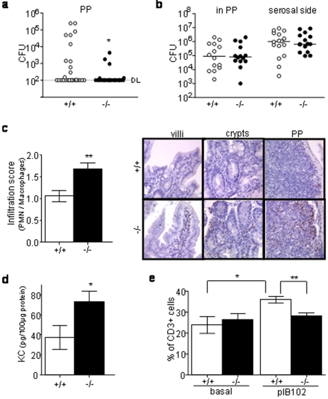
Figure 3. Nod2 modulates the local intestinal inflammatory response during Y. pseudotuberculosis infection.
(a, c–d) Nod2−/− and Nod+/+mice were orogastrically inoculated with 1×10⁷ CFU of YPIII(pIB102) bacteria and sacrificed 5 days after inoculation. Their PP were removed and analyzed. (a) Bacterial counts were significantly lower in Nod2−/− (n = 29) than in Nod+/+ (n = 27) mice (Mann-Whitney-test) for a detection limit (DL) of 102 cfu. (b) PPs from non infected mice were placed in a Ussing chamber and 1×10⁸ CFU/ml pIB102 were placed in the mucosal compartment of the chamber. No differences between Nod2−/− (n = 7) and Nod2+/+ (n = 7) mice for the bacterial translocation trough PP (mucosal to serosal flux, P = 0.85) as well as for the bacterial colonisation of the tissue (count of the bacteria remaining in the tissue after the experiment; P = 0.94) were seen at 120 min (Mann-Whitney test). Detection limit was 102 CFU. (c) Infiltration of macrophages and neutrophils was scored in PPs and their surrounding epithelium. The infiltration score was higher in Nod2−/− (n = 9) than in Nod2+/+ (n = 11) mice (Student t-test). Photos show representative MPO staining of intestinal villi, crypts and PPs (d) KC concentrations were recorded by ELISA. KC level was higher in Nod2−/− (n = 6) than in Nod2+/+ (n = 7) mice (Student t-test). (e) Flow cytometry analyses revealed an increase of the relative proportion of CD3+ T-cells after infection in Nod2+/+ mice (n = 6) but not in Nod2−/− mice (n = 6) (Student t-test). Error bars indicate mean+/−SEM. *P<0.05, **P<0.01, ***P<0.001.

