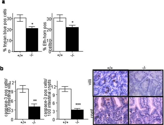
Figure 4. Less apoptosis of epithelial cells surrouding PP areas in Nod2−/− mice.
Nod2−/− and Nod+/+mice were orogastrically inoculated with 1×10⁷ CFU of YPIII(pIB102) bacteria and sacrificed 5 days after inoculation. Their intestines were removed and analyzed.(a) Cell death measured by the proportion of Trypan blue or Ethidium homodimer-1 positive cells was lower in the PPs of Nod2−/− mice (Student t-test). (b) Apoptosis measured by the number of caspase-3 stained epithelial cells in the 100 intestinal villi and crypts surrounding PPs was also lower in Nod2−/− mice (Student t-test). Photos show representative caspase-3 staining of intestinal villi and crypts. Error bars indicate mean+/−SEM. *P<0.05, **P<0.01, ***P<0.001.

