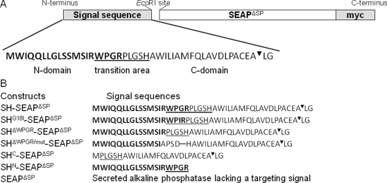
Figure 2. Shrew-1 (SH) signal sequence and the construction of the SEAP fusion proteins.
(A) Organization of shrew-1 signal sequence. Bold: N-domain (shrew-1 residues 1–19). Standard type: C-domain (shrew-1 residues 20–43). Underlined: transition area (shrew-1 residues 16–24). ▾: signal sequence cleavage site. LG: shrew-1 residues 44 and 45. (B) Diagrams of SEAP constructs with assigned shrew-1 signal sequences. Signal sequences are N-terminally fused to the SEAP protein lacking the endogenous signal peptide (SEAPΔSP). C-terminally, all fusion proteins are tagged with myc (EQKLISEEDL). For cleavage site recognition (PACEA▾LG) shrew-1 residues 44 and 45 (LG) are included in the constructs.

