Figure 4.
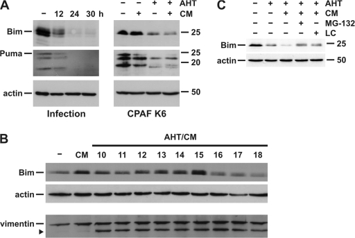
BH3-only proteins are degraded by infection with C. trachomatis or upon prolonged expression of active CPAF. (A) (Left) T-REx-293 cells were infected with C. trachomatis for indicated time points and RIPA buffer extracts were prepared. (Right) CPAF K6 cells were treated with either 5 ng/μl AHT, CM, or both as indicated. Cell extracts were analyzed by Western blotting using antibodies specific for Bim, Puma, or actin as loading control. (B) Time course of cleavage of CPAF substrates. CPAF K6 cells were treated with 5 ng/μl AHT and CM to induce CPAF expression for the indicated time periods. Cell extracts were analyzed by Western blotting. The arrowheads indicate cleavage product of vimentin. (C) Inhibition of Bim degradation by proteasome inhibitors. CPAF K6 cells were treated with 5 ng/ml AHT or 5 ng/ml AHT plus CM. 6 h before cell harvesting, either 40 μM MG-132 or 5 μM clasto-lactacystin β-lactone (LC) were added. Cell extracts were analyzed by Western blotting.

