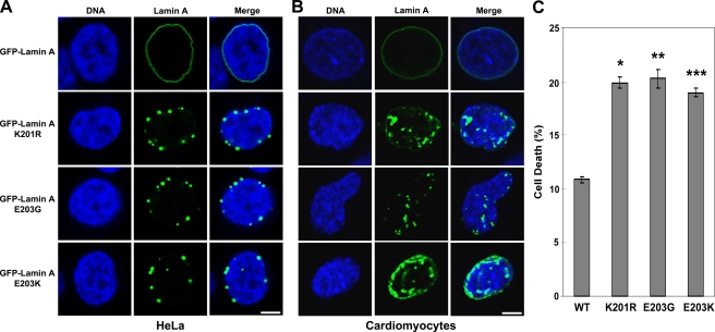
Figure 3.
SUMO site K201R mutant lamin A and cardiomyopathy-associated E203G and E203K mutant lamin A proteins exhibit similar patterns of aberrant localization. (A and B) Wild-type, K201R, E203G, and E203K lamin A GFP fusion expression plasmids were transfected into HeLa cells (A) or mouse embryonic cardiomyocytes (B), and the subcellular localization of the GFP–lamin A proteins was examined by confocal fluorescence microscopy. DNA was visualized by staining with HOECHST 33342. Bars, 5 μm. (C) Wild-type, K201R, E203G, or E203K lamin A GFP fusion expression plasmids were transfected into HeLa cells, which were then analyzed by trypan blue assay to measure cell death. Data are shown as means ± SEM. (*, P < 0.0001; **, P < 0.0002; ***, P < 0.0001 [for lamin A mutants vs. wild type]) and are each from three datasets.

