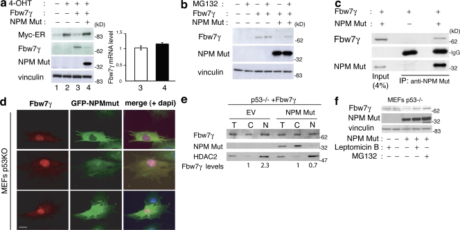
Figure 5.
Mutant NPM delocalizes and destabilizes Fbw7γ. (a, left) WB analysis in p53−/−;Myc-ER cells infected with empty (No. 2) or HA-Fbw7γ-expressing (No. 4) retroviruses. HA-Fbw7γ–infected cells were reinfected with empty or NPMmut-expressing lentiviruses (also expressing GFP as marker). Cells were treated for 2 h with 4-OHT (Nos. 2–4). (right) QPCR analysis of Fbw7γ mRNA levels in samples Nos. 3 and 4. Data represent the mean of three determinations ± SEM. (b) WB analysis in the same cells as in panel a. Cells were or were not treated with MG132 for 2 h, as indicated. (c) dKO MEFs were cotransfected with plasmids expressing flag-Fbw7γ and NPMmut or the empty vector, as indicated. Total lysates and IPs were blotted with antibodies against NPMmut or the flag epitope. (d) IF analysis of p53−/− MEFs infected with GFP-NPMmut– and flag-Fbw7γ (red staining)-expressing constructs. A merge of the NPMmut, Fbw7γ, and DAPI staining is also shown. Bar, 10 μm. (e) Nucleus–cytoplasm fractionation in p53−/− MEFs expressing flag-Fbw7γ infected with GFP-NPMmut or control (EV) retroviruses. Total cell lysates (T) or cellular fractions (C, cytoplasm; N, nucleus) were analyzed by WB as indicated. (f) The same cells as in panel e were or were not treated with 1 μM LMB or MG132 for 3 h, as indicated. Expression of Fbw7γ was analyzed by WB on total cells lysates using anti-flag antibodies.

