Figure 8.
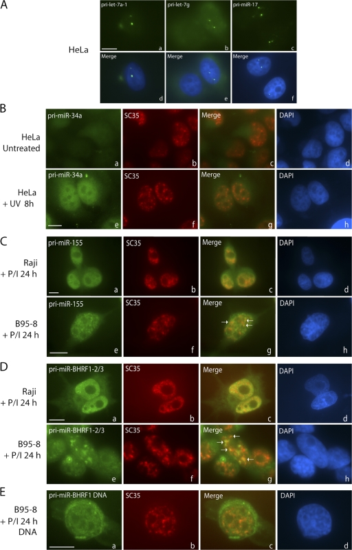
Localization of endogenous pri-miRNAs. (A) Pri-miRNAs expressed at low levels were detected using amplification and localize to one or two foci per cell. (B) Pri-miR-34a is not detected in untreated HeLa cells (a–d) but shows diffuse nucleoplasmic localization in HeLa cells after UV irradiation (e–h). Costaining for SC35 is shown in panels b and f. (C) BIC/pri-miR-155 localizes throughout the nucleoplasm in Raji (a–d) and B95-8 cells treated with PMA and ionomycin (P/I) for 24 h, as well as in nuclear foci in B95-8 cells (e–h), many of which overlap with SC35 (g, arrows). (D) Pri-miR-BHRF1-2∼3 shows diffuse nucleoplasmic and cytoplasmic distribution in Raji cells (a–d) but is concentrated in nuclear foci in B95-8 cells (e–h), some of which overlap with SC35 (g, arrows). (E) EBV BHRF1 DNA localizes diffusely in the nucleus and cytoplasm (due to viral release). Bars, 10 μm.

