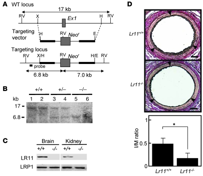
Figure 1. Intimal thickness of arteries after cuff placement in Lr11–/– mice.
(A) Targeted disruption strategy of the murine LR11 gene, consisting of 49 exons. The targeting vector (bold line) contains 3.3 kb (5′) and 4.4 kb (3′) of genomic DNA flanking the neomycin-resistance cassette (Neor). After homologous recombination, Neor replaced exon 1 (Ex1, gray box), which contained the initiation codon of the LR11 gene. The location of the probe used for Southern blot analysis is shown. RV, EcoRV; X, XbaI; H, HindIII; E, EcoRI. (B) Southern blot analysis of murine-tail DNA from heterozygous intercrosses digested with EcoRV using the probe (see Figure 1A) that detects 17-kb and 6.8-kb fragments in the WT and knockout allele, respectively. (C) Immunodetection of LR11 protein. Total protein (100 μg) extracted from brain and kidney were separated by electrophoresis, blotted on a membrane, and incubated with antibody against LR11 (~250 kDa) or LRP1(~85 kDa). The samples were loaded on the same gel but not on immediately neighboring lanes. (D) Upper panels show sections of femoral artery of Lr11+/+ or Lr11–/– mouse after cuff placement, subjected to elastica van Gieson staining. Arrowheads indicate the internal elastic layers. Scale bar: 50 μm. Lower panel shows I/M ratio of arteries presented as mean ± SD (n = 15). *P < 0.05.

