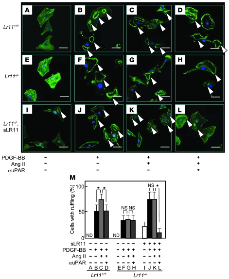
Figure 4. Ang II–induced membrane ruffle formation of cultured SMCs derived from Lr11–/– mice.
(A–L) Ang II–induced membrane ruffling (arrowheads) in Lr11+/+ or Lr11–/– SMCs. The indicated SMCs were incubated with or without Ang II (1 μM) for 24 hours in the presence or absence of recombinant sLR11 (1 μg/ml) with or without anti-uPAR antibody for 24 hours. PDGF-BB (10 ng/ml) was then added to the culture medium for 10 minutes before immunofluorescence analyses. Cells were then stained using Alexa Fluor 488 phalloidin. Scale bars: 10 μm. (M) The number of cells with membrane ruffles were counted among 500 cells in the field. Data are presented as mean ± SD (n = 3). *P < 0.05. ND, not detected.

