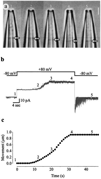
Figure 2.
Depolarization-induced displacement of the membrane patch is associated with activation of MS channels. (a) High-resolution video microscopy of a patch of membrane in a patch pipette. The membrane patch (arrows) moves into the pipette during depolarization. (Bar = 2 μm.) (b) After a voltage step from −80 to +80 mV (Upper trace), the nine MS channels in the membrane patch start to open after a delay of ≈10 s (Lower trace), with full activation at ≈20 s. When the voltage is stepped back to −80 mV, the MS channels then deactivate over a period of ≈10 s. Channel opening is indicated by upward current steps at positive membrane potentials and downward current steps at negative membrane potentials. The leakage current through the patch, indicated by the 18-pA current steps that coincide with the voltage steps, was not subtracted from the current record. (c) Plot of the distance moved by the membrane patch vs. time after the voltage step. The corresponding numbers 1–5 in the three parts of the figure indicate the time points for simultaneous measurement of video and current data from the same inside-out patch excised from a Xenopus oocyte with a borosilicate glass pipette.

