Figure 2.
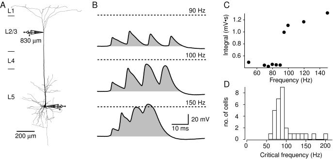
Voltage integral as a measure of critical frequency across neurons. (A) Reconstruction of a biocytin-filled L5 pyramidal neuron from a 57-day-old rat with a dendritic whole-cell patch pipette located 830 μm from the soma and a second pipette located at the soma. (B) Four 5-ms square-pulse current injections at the soma at 90 Hz (top trace) elicited APs at the soma that had amplitudes of approximately 20–30 mV recorded at the dendritic site. The shaded region represents the voltage time integral, which had a value of 0.50 mV⋅s. At the critical frequency, 100 Hz (middle trace), the integral increased to 1.28 mV⋅s. At 150 Hz (bottom trace), the Vm crossed 0 mV, but the integral did not greatly increase. The dashed lines represent 0 mV. (C) The integral of four successive APs over time at different frequencies in the same neuron shows a sharp nonlinear increase at the critical frequency. (D) The critical frequencies of 31 different L5 pyramidal neurons. The average value was 98 ± 6 Hz, but the distribution was skewed toward higher values. In two cells, no detectable critical frequency was reached below 200 Hz (data not shown).

