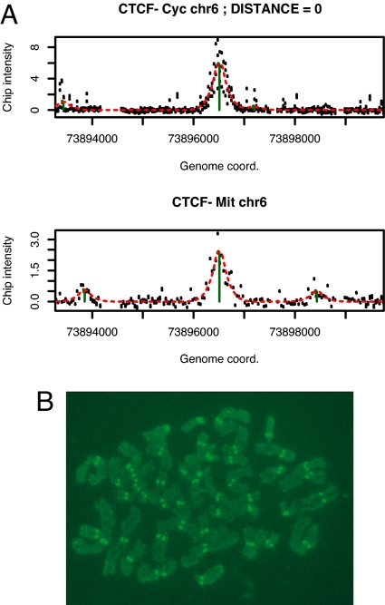
Fig. 6.
Association of CTCF with chromosomal sites and centromeres during mitosis. (A) Example of a CTCF site on chromosome 6 occupied in both asynchronously growing cells (Upper, CTCF Cyc) and cells arrested in mitosis (Lower, CTCF-Mit). The calculated distance between the peaks is indicated (distance = 0). A complete list of all sites detected in both asynchronously growing and mitotic cells is shown in Table S4. (B) Immunofluorescence detection of CTCF bound to centromeres of human mitotic chromosomes. A representative example of a mitotic chromosome preparation is shown. All cells examined from each preparation showed centromeric staining by CTCF.

