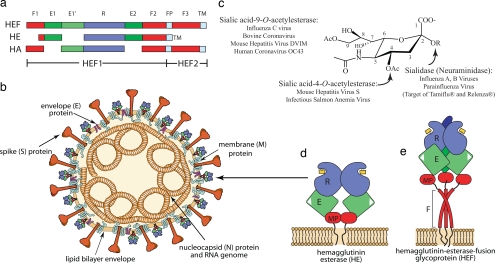
Fig. 1.
Comparison of the structural and functional activities of HE, HEF, and HA. (a) Linear order of the sequence segments in HEF, HE, and HA color-coded by domains. Red segments, F1, F2, and F3; green, E1 and E2; lighter green, E′; blue, R; light blue, fusion peptide (FP) or transmembrane domain (TM). The HEF subunit 1 (HEF1) and subunit 2 (HEF2) are also indicated. HEF2 is absent from HE. This figure is adapted from refs. 1, 3, and 5. (b) Illustration of a group 2a coronavirus with the indicated virion proteins. Individual hemagglutinin molecules (HE) are color coded as described for a. (c) Substrate specificity of viral HE, HEN, and NA toward SAs. Figure is adapted from ref. 3. (d) Schematic illustration of the HE dimer. Colors are as described for a. The membrane-proximal domain (MP) is also shown. The catalytic triad is represented in the E domain as a white triangle. Binding of SA to the R domain is shown as a yellow box. The TM domain is shown embedded in the virion envelope. (e) Schematic illustration of the HEF trimer. The F domain comprising F1, F2, F3, and FP that forms a coiled-coil is indicated. Other features are as described for d.

