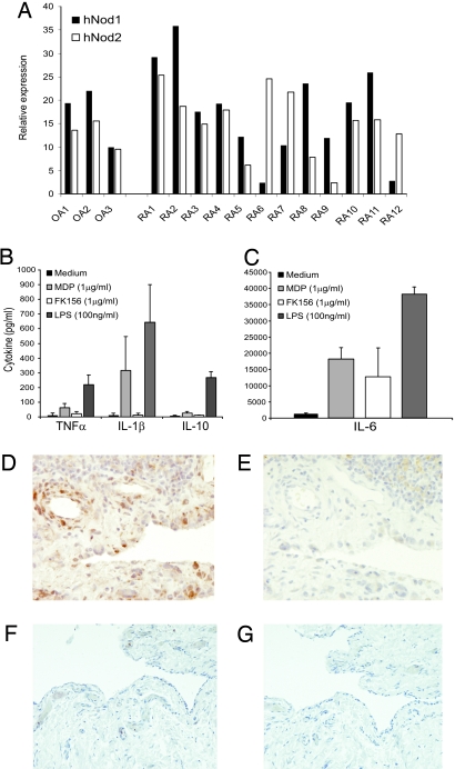
Fig. 1.
Nod expression in RA synovial tissue. (A) mRNA expression of Nod1 or Nod2 in RA and OA synovial tissue samples expressed as relative expression to the houskeeping gene GAPDH (2−delta Ct value × 1,000). (B and C) Cytokine production of RA synovial tissue after exposure to MDP, FK156, and LPS. (D) MDP-containing fragments in RA synovial tissue showed by immunohistochemical staining of paraffin sections using MoAb 2–4-5 (39). (E) Isotype control. Both synovial lining cells and endothelial cells contain MDP fragments. (D and E) ×200 magnification. (F) MDP staining of OA synovial tissue; note almost no staining. Isotype control. (F and G) ×100 magnification.

