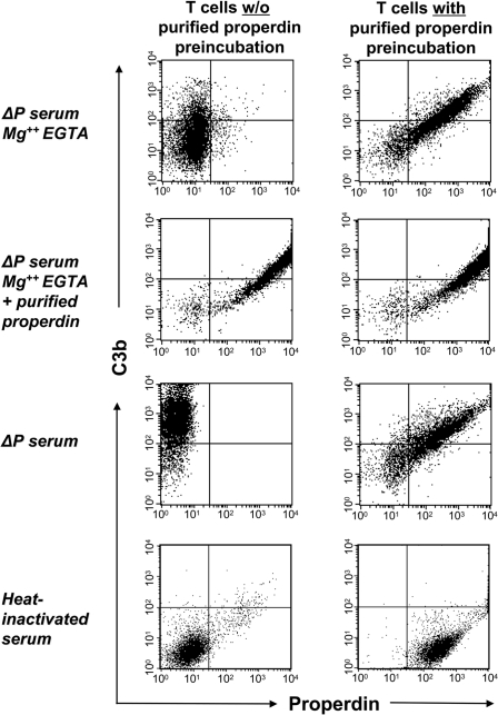
Fig. 2.
Properdin binding to apoptotic T cells initiates C3b deposition. Apoptotic T cells were either left untreated (Left) or preincubated with 5 μg/ml purified properdin (Right). Cells were washed and then incubated for 15 min with 15% properdin-deficient (ΔP) serum diluted in Mg2+ EGTA, ΔP serum diluted in Mg2+ EGTA and reconstituted with purified properdin, 15% ΔP serum diluted in PBS, or 15% heat-inactivated human serum diluted in PBS. Deposition of C3b on apoptotic T cells was measured by FACS analysis (with the following MFIs: uppermost panel in Left, 35 ± 5; uppermost panel in Right, 320 ± 45; second panel in Left, 720 ± 25; second panel in Right, 710 ± 40; third panel in Left, 850 ± 50; third panel in Right, 420 ± 40; lowermost panels in Left and Right, <10). Gates were set at the complete apoptotic cell population, excluding only viable cells. Results shown are representative of three independent experiments.

