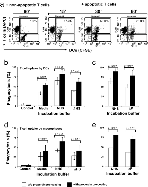
Fig. 3.
Quantification of properdin-mediated uptake of apoptotic T cells by macrophages and DCs. (a) FACS-based assay for the measurement of ingested T cells. DCs were grown to confluence and labeled with CFSE. Apoptotic T cells were labeled with an APC-conjugated anti-CD4 mAb and added to the DCs in media containing 15% NHS. At indicated time points, nonbound/ingested T cells were aspirated and DCs were detached. The percentage of CFSE/APC-positive DCs (CFSE-labeled DCs that ingested apoptotic APC-labeled T cells during the incubation period) was determined by FACS analysis and is shown within the boxes. (b and c) Properdin increases uptake of apoptotic T cells by DCs. Apoptotic, APC-labeled T cells were either incubated with purified properdin (5 μg/ml media) or left untreated. T cells were washed and then incubated with DCs in media, media with NHS, media with ΔHS (b), or media with properdin-depleted serum (ΔP) (c) for 60 min, and T cell uptake was measured by FACS. (d and e) Properdin increases uptake of apoptotic T cells by macrophages. Experiments were performed as in b and c but with macrophages in place of DCs. Shown are the mean ± SD values of three separate experiments. Statistical difference between the ingestion of T cells either with or without properdin preincubation by DCs and macrophages was P < 0.01 in all cases.

