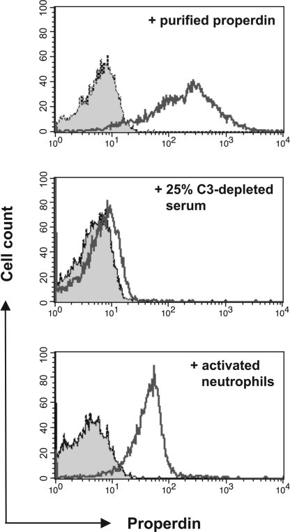
Fig. 5.
Neutrophil-derived properdin binds to apoptotic T cells. (Top) FACS analysis of properdin bound to apoptotic T cells after their incubation with purified properdin. (Middle) Measurement of bound properdin on apoptotic T cells after their incubation for 15 min with 25% C3-depleted human serum diluted with PBS. (Bottom) Analysis of properdin bound to apoptotic T cells after their incubation with activated neutrophils. TNF-α (100 nM) was added to freshly isolated neutrophils mixed with apoptotic T cells, and cell mixtures were incubated for 15 min at 37°C in RPMI medium 1640. Cell mixtures were analyzed for deposition of properdin on T cells by FACS. The shaded histograms depict staining with the isotype control mAb. Bound properdin is depicted by the nonshaded histograms. All datasets shown are one representative result of at least three independently performed experiments. Activation of neutrophils with PMA or GM-CSF resulted in comparable amounts of properdin deposition on apoptotic T cells (data not shown).

