Fig. 5.
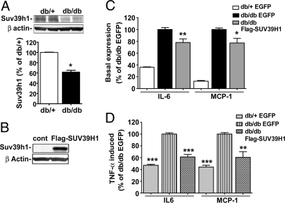
Suv39h1 levels in VSMC regulate inflammatory gene expression. (A) Western blot analysis of cell lysates from db/+ and db/db VSMC using SUV39H1 and β-actin antibodies. The bar graph represents mouse Suv39h1 protein levels by densitometry and are expressed as the percentage of db/+ VSMC (mean ± SE; *, P < 0.023 vs. db/+, n = 3). (B) Western blot analysis of cell lysates from db/db VSMC transfected with EGFP or FLAG-SUV39H1 vectors by using SUV39H1 and β-actin antibodies. (C and D) VSMC were transfected with EGFP or FLAG-SUV39H1 vectors and inflammatory gene levels measured in basal or TNF-α-treated cells (10 ng/ml for 1 h). RT-qPCR results shown as the percentage of db/db-EGFP. (C) Basal gene expression. Data represent mean ± SE of triplicate qPCRs from two independent experiments (*, P < 0.05; **, P < 0.01 vs. db/db-EGFP). (D) TNF-α-induced gene expression. Results represent mean ± SE of triplicate qPCRs, n = 2 for IL-6 and n = 3 for MCP-1 (**, P < 0.01; ***, P < 0.0001 vs. db/db-EGFP).

