Fig. 2.
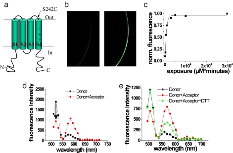
Associated VSOP subunits in the plasma membrane. (a) VSOP construct containing S242C mutation. (b) Confocal images of oocyte expressing WT Ci-VSOP (Left) and 242C Ci-VSOP (Right) channels labeled with Alexa488-maleimide. (Magnification: × 50.) (c) Labeling time course for Alexa488-maleimide of 242C fitted with an exponential function with τ = 670.5 μM × min. (d) Representative fluorescence spectrum from an oocyte expressing Ci-VSOP S242C channels first labeled with donor fluorophore only (Alexafluor488-maleimide, ≈20% of the subunits labeled; squares) and then labeled to saturation with acceptor fluorophore (TMR-MTS; circles). FRET efficiency was measured as the % decrease in donor fluorescence (arrow). (e) Representative fluorescence spectrum from an oocyte expressing Ci-VSOP S242C channels first labeled with donor fluorophore only (Alexafluor488-maleimide, ≈20% of the subunits labeled; squares), labeled to saturation with acceptor fluorophore (TMR-MTS; circles), and exposed to DTT to restore the donor fluorescence (10 mM DTT for 10 min; triangles).

