
Figure 2.
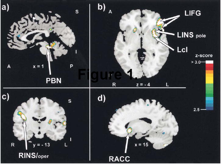
Increases in regional cerebral blood flow during stomach distension. Arrows point to significant changes in blood flow in the difference image (inflation minus deflation). Letters and numbers below the slices indicate their orientation and stereotactic coordinates. A = anterior; I = inferior; L = left; Lcl = left claustrum; LIFG = left inferior frontal gyrus; LINS = left insula; LINS pole = left insular pole; P = posterior; PBN = parabrachial nucleus; R = right; RACC = right anterior cingulate cortex; rCBF = regional cerebral blood flow; RINS/oper = right insula/operculum; S = superior; x = sagittal plane; y = coronal plane; z = horizontal plane.