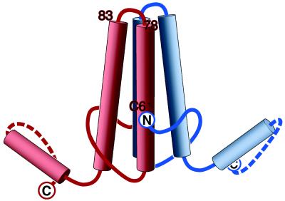
Figure 5.
Model of the HBV capsid protein dimer. The locations of the amino acids that have now been localized are marked: (i) the N terminus (this study), shown with the polypeptide chain at this point oriented as suggested by our difference maps (Fig. 4); (ii) the C terminus (5); and (iii) the loop covering residues 78–83 (7). The position of Cys 61 has also been inferred (Discussion). In most respects, this model confirms and supports that of Böttcher et al. (1), but has been revised in light of more recent information. The handedness, originally assigned arbitrarily (1), has been switched on the basis of an experimental determination (7). There are also minor differences in the positions assigned to residue 149, which we assign to the inner surface with the chain direction as shown, and the N terminus, which we place ≈10 Å farther up from the bottom of the molecule, as shown.

