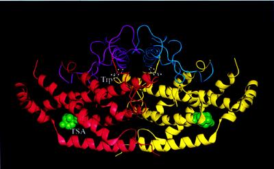
Figure 1.
Schematic representation of the dimeric structure of YCM. The allosteric domain of the left monomer (in red) is drawn in magenta and that of the right monomer (in yellow) is drawn in blue. The two effector tryptophans (in white) are in ball-and-stick form and the transition state analogues (in green) are in space-filling model. This figure, as well as Figs. 4–7 and 9, was made with quanta (Molecular Simulations, San Diego, CA) [(modified from (4)].

