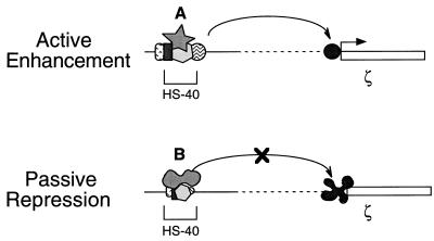
Figure 7.
A cartoon model of the HS-40 function. It is proposed that the multiple protein–DNA complex(es) formed within HS-40 adopts different conformations in the embryonic and adult erythroid cells. This conformational difference results at least in part from competitive binding between different nuclear factors, A and B, at the 3′NF-E2/AP1 motif. In the upper scheme, the HS-40 enhancer would activate transcription of the ζ-globin gene through interaction with its promoter. On the other hand, the altered HS-40 complex shown in the lower scheme is unable to overcome the negative regulatory effect of repressing elements in the ζ-globin promoter. See text for more discussion.

